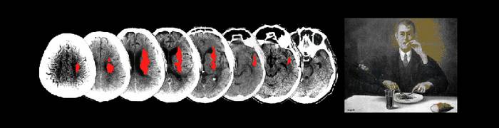


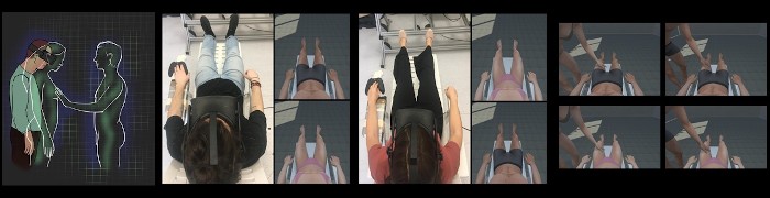
Monkey neurophysiology and human neuroimaging studies have demonstrated that passive viewing of optic flow stimuli activates a cortical network of temporal, parietal, insular, and cingulate visual motion regions. Here, we tested whether the human visual motion areas involved in processing optic flow signals simulating self-motion are also activated by active lower limb movements, and hence are likely involved in guiding human locomotion. To this aim, we used a combined approach of task-evoked activity and resting-state functional connectivity by fMRI. We localized a set of six egomotion-responsive visual areas (V6+, V3A, intraparietal motion/ventral intraparietal [IPSmot/VIP], cingulate sulcus visual area [CSv], posterior cingulate sulcus area [pCi], posterior insular cortex [PIC]) by using optic flow. We tested their response to a motor task implying long-range active leg movements. Results revealed that, among these visually defined areas, CSv, pCi, and PIC responded to leg movements (visuomotor areas), while V6+, V3A, and IPSmot/VIP did not (visual areas). Functional connectivity analysis showed that visuomotor areas are connected to the cingulate motor areas, the supplementary motor area, and notably to the medial portion of the somatosensory cortex, which represents legs and feet. We suggest that CSv, pCi, and PIC perform the visual analysis of egomotion-like signals to provide sensory information to the motor system with the aim of guiding locomotion.
![]()
![]()
![]()

Hum Brain Mapp. 2019 Aug 1;40(11):3174-3191. doi: 10.1002/hbm.24589
